
MelaNailScan
- Subungual Melanoma Diagnostic Support Software -


What is the disease?
What does it do?
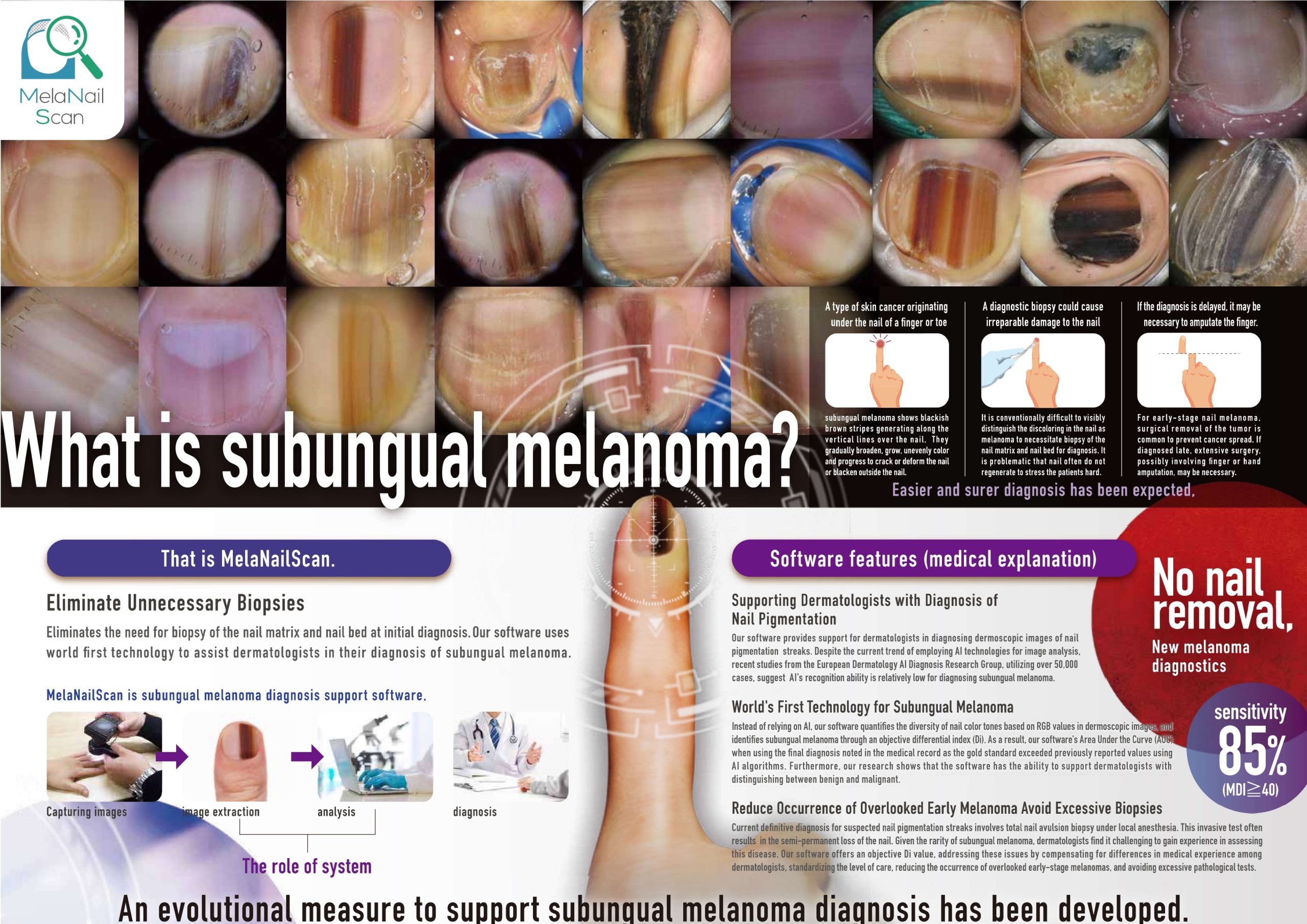
Subungual melanoma, a form of skin cancer originating in the nail unit,
is a rare and difficult condition to diagnose. Our software, developed in
Japan and the first of its kind in the world, is designed to aid doctors in
identifying this illness.
Due to the rarity of the disease, few doctors are equipped to diagnose
it, and traditional diagnostic methods require removing the affected nail,
which may not regrow afterwards. Additionally, the disease is not widely
recognized globally. Late detection can lead to severe mental and physical
burden for patients, which can ultimately lead to amputation of a digit.
What kind of examination
does it provide?

Software features
(medical explanation)

Sales/Service base
Q'sfix Corporation
Headquarters: Marunouchi 1-chome 7-12 Sapia Tower 26F,
Chiyoda-ku, Tokyo, 100-0005
TEL: +81-3-6256-0330
Production base
Human Engineering Co.,Ltd.
Shizuoka Headquarters: 166-3 Araya, Mishima-shi,
Shizuoka, 411-0834
TEL:+81-55-983-3877
E-mail:melanailscanloajrhgp@human.co.jp